
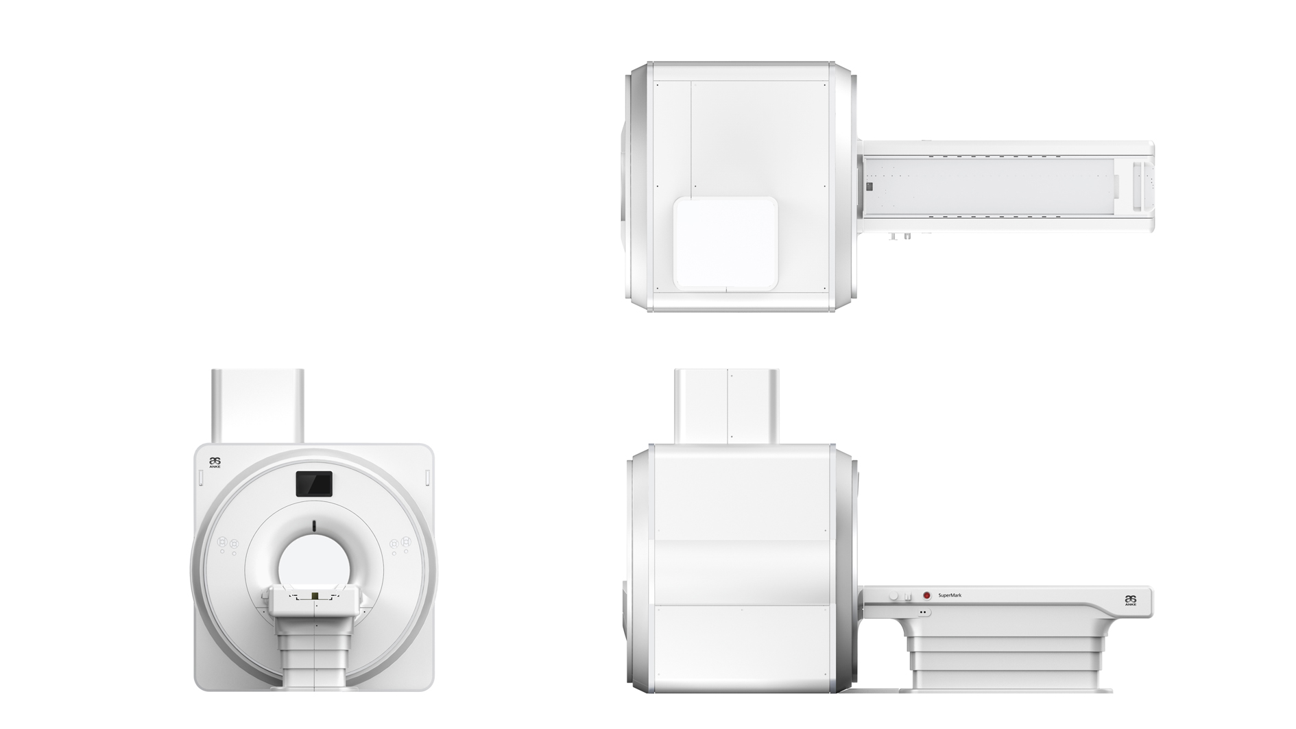
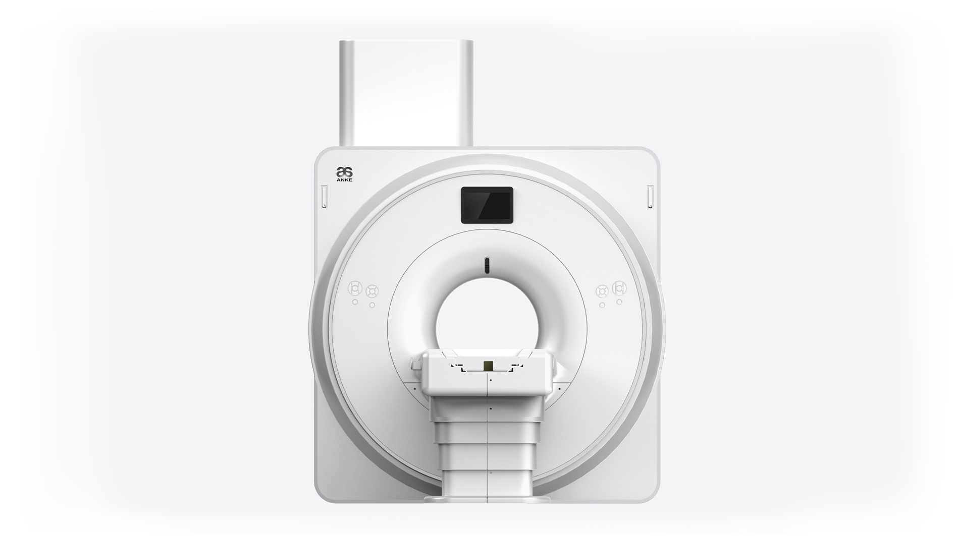
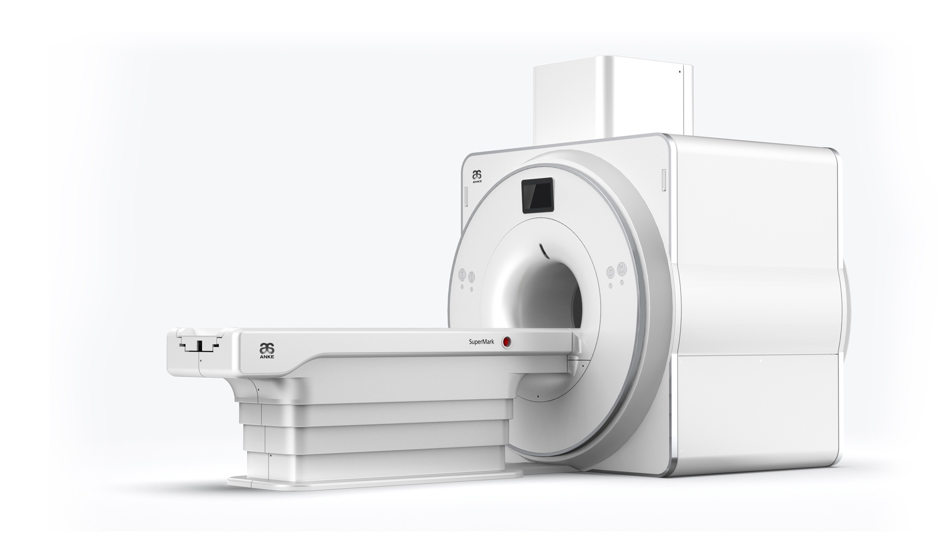
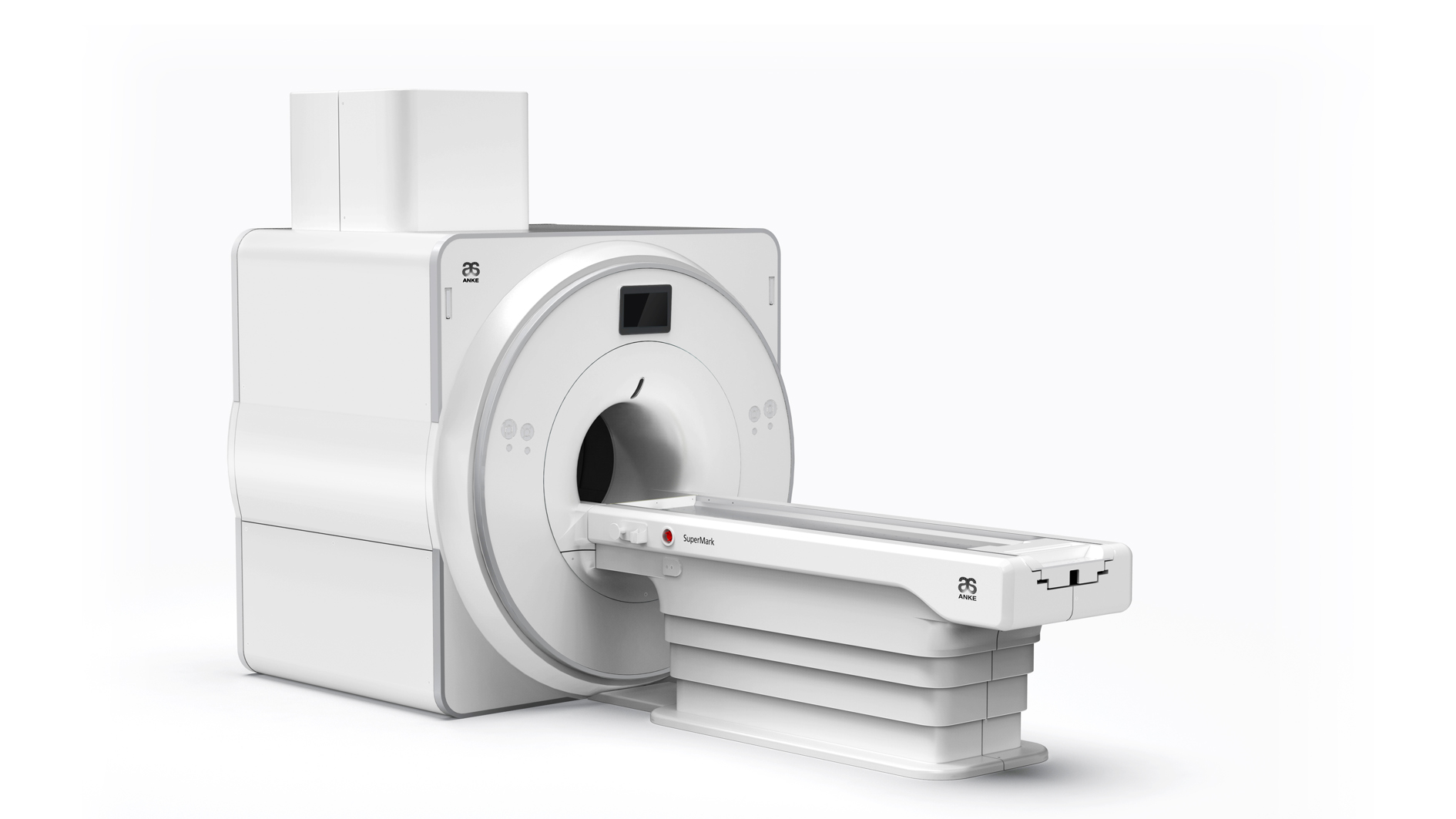
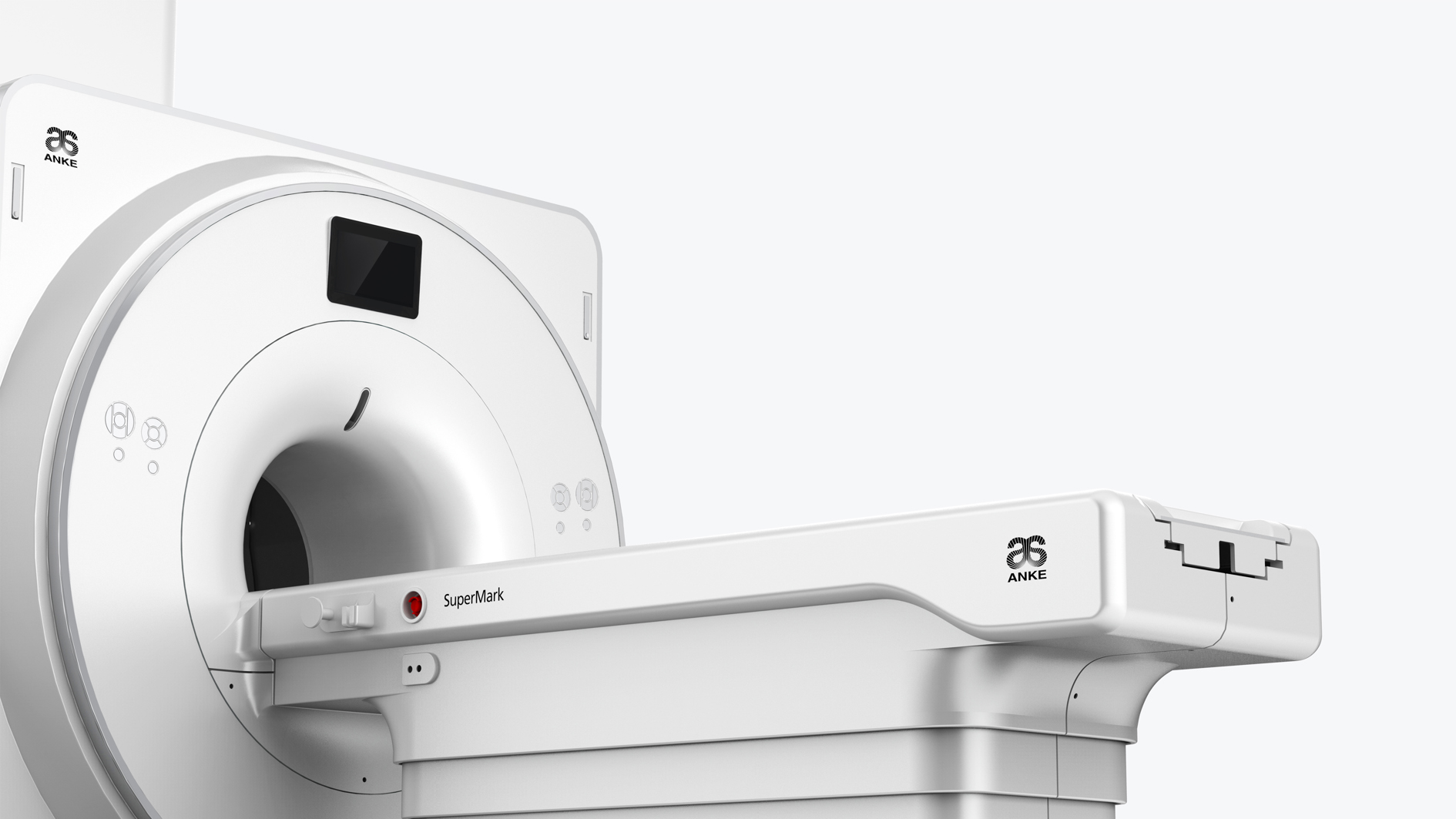
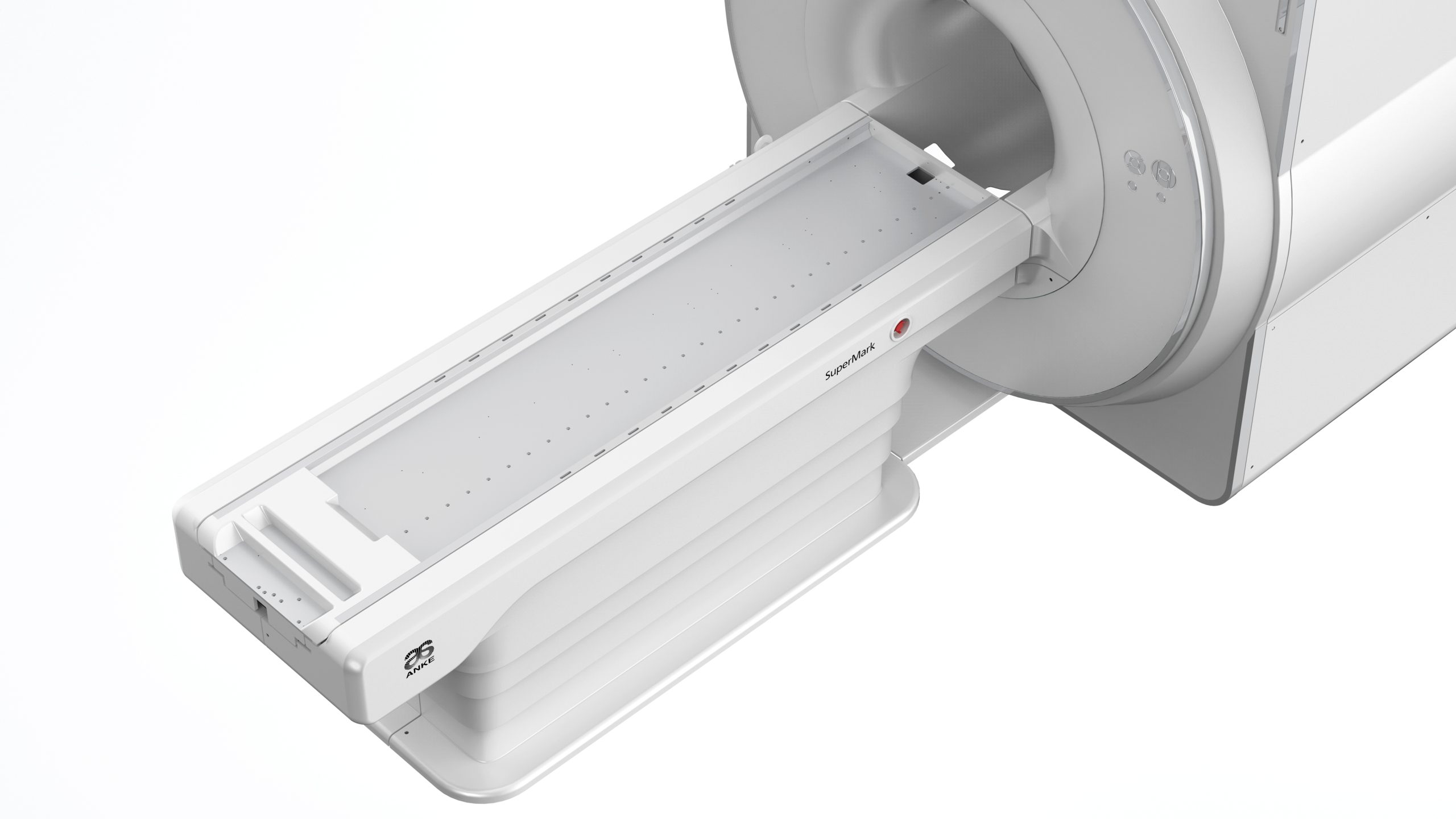
SuperMark 1.5T is a modern superconducting MRI system from ANKE, developed on the basis of many years of experience in the field of research and production of medical equipment. The system is designed for full-fledged magnetic resonance examination of the whole body, including:
- nervous system;
- soft tissues of the joints;
- abdominal and pelvic organs.
The main features of SuperMark 1.5T:
- It supports both traditional clinical diagnostics and advanced technologies, including 3D angiography;
- It is equipped with the innovative ANKE APEX operating system, which provides convenient and intuitive operation;
- Ensures fast image processing, which increases research efficiency.
The SuperMark 1.5T system is an ideal solution for medical institutions seeking to combine diagnostic accuracy and modern technology.
Technical characteristics of MRI SuperMark 1.5T:
Magnet type – superconducting
Magnetic field strength – 1.5T
Magnetic field stability ≤ 0.1 ppm/h
Magnetic field homogeneity (VRMS measurement method):
- 50 cm DSV (VRMS) ≤ 0.800 ppm
- 45 cm DSV (VRMS) ≤ 0.450 ppm
- 40 cm DSV (VRMS) ≤ 0.170 ppm
- 30 cm DSV (VRMS) ≤ 0.058 ppm
- 20 cm DSV (VRMS) ≤ 0.020 ppm
- 10 cm DSV (VRMS) ≤ 0.002 ppm
Net weight of the magnet (including the weight of liquid helium) – 3800 kg
Volume of liquid helium (100% filling) – 700 liters
Zero evaporation technology of liquid helium
The length of the magnet:
- The length of the magnet tunnel (without the body) is 157 cm;
- The length of the magnet tunnel (with the body) is 170 cm.
Magnet bore diameter – 605 mm ± 5 mm
Maximum open aperture of the magnet (at both ends) – 160 cm
Patient table, maximum width – 46 cm
Forming images
- 256 x 256 – maximum refresh rate (FFT) ≥ 3300 frames per second
- 2D – thickness 0.1 mm
- 3D – thickness 0.05 mm
- Maximum FOV – 50 cm
- Minimum FOV – 1 cm
Gradient system
- Max. amplitude (uniaxial, ineffective value) – 40 mT/m
- Max. ramp-up speed (single axis, ineffective value) -150 T/m/s
- Rise time – 0.27 ms
Radio frequency system
- RF amplifier with a maximum power of 20 kW
- The center frequency is 63.85 MHz
- Number of receiving and transmitting channels – 16
- The maximum resolution of the received signal is 16 bits
- Sample resolution – 100 ns
Multi-channel phased receiver coils
- Head-neck coil (16 channels)
- Flexible coil for the whole body (16 channels)
- Knee coil (8 channels)
- Shoulder coil (4 channels)
- Head coil (8 channels) (optional)
- Neck coil (8 channels) (optional)
- Coil for hand, wrist (8 channels) (optional)
- Coil for lower leg, foot (8 channels) (optional)
- Chest coil (8 channels) (optional)
Electric patient table
- Table length – 260 cm
- Horizontal movement range – 185 cm
- Minimum height – 63 cm
- Vertical range of motion – 28 cm
- Maximum patient weight – 200 kg
- Maximum horizontal movement speed ≥ 20 cm/s
- Position control system is located on both sides of the table
- Lighting, ventilation, call system in the magnet tunnel
Workstation
- Windows-based computer operating system
- CPU – 3.6 GHz (i7, eight cores)
- RAM – 16 GB
- Hard disk – 1 TB x 2
- Number of saved images (256 x 256 matrix) – ≥ 2,750,000
- Display size – 24″
- External storage of image data – DVD/USB
- Full compatibility with DICOM 3.0
Scanning technologies and sequences
- Spin echo (SE), SE T1 weighted image, SE proton density (PD) weighted image, SE dual contrast, 3D SE, SE sequence off
- Fast Subtraction (FSE), FSE T2-weighted image, FSE T2-weighted image, FSE proton density, weighted image, 3D FSE and Subtraction FSE
- Inversion recovery (IR), IR fat suppression, IR water suppression, IR silencing sequence
- Fast inversion recovery (FIR), fat suppression FIR, water suppression FIR, fat suppression STIR, FIR
- Mute the sequence
- Gradient echo (GRE), GRE T1-weighted image, GRE T2-weighted image, rapidly reoriented GRE, GRE breath-hold
- 3D fast GRE, 3D fast reoriented GRE, phase/anti-phase GRE
- Balanced stationary free process (B-SSFP) GRE
- Magnetic resonance angiography (MRA)
- TOF 2D & 3D, PC, CE-MRA
- Advanced imaging software, diffusion weighted imaging (DWI), head DWI, abdomen DWI, chest DWI, prostate DWI
- Cholangiopancreatography (MRCP), magnetic resonance urography (MRU), magnetic resonance myelography (MRM)
Visualization technologies
- Fast visualization technology
- Dual-engine parallel scintigraphy technology, ultrafast SSFSE imaging
- Artifact suppression technology
- Pre-saturation technology, flow compensation technology, screw scanning technology, lattice technology
- Magnetic susceptibility images (SWI)
- Image evaluation software
- Image processing software
- Multidimensional reconstruction MPR, reconstruction MIP, reconstruction MinIP
- Software for system management
- Control software, calibration software
- Diffusion tensor imaging (DTI)
- Heart cat
- Dynamic visualization technology
- Dynamic enhancement FAST 3D, dynamic enhancement FAST 4D
- 2D, 3D, multi-layer scanning technology
- Sequence of noise reduction pulses
- Accelerated scan sequences
- Special methods for filling k-space and processing data
- Propeller motion artifact reduction technology
- DWI diffusion-weighted studies
