
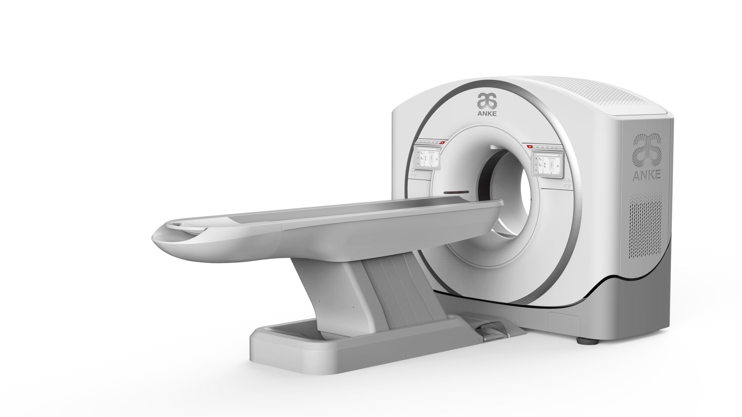
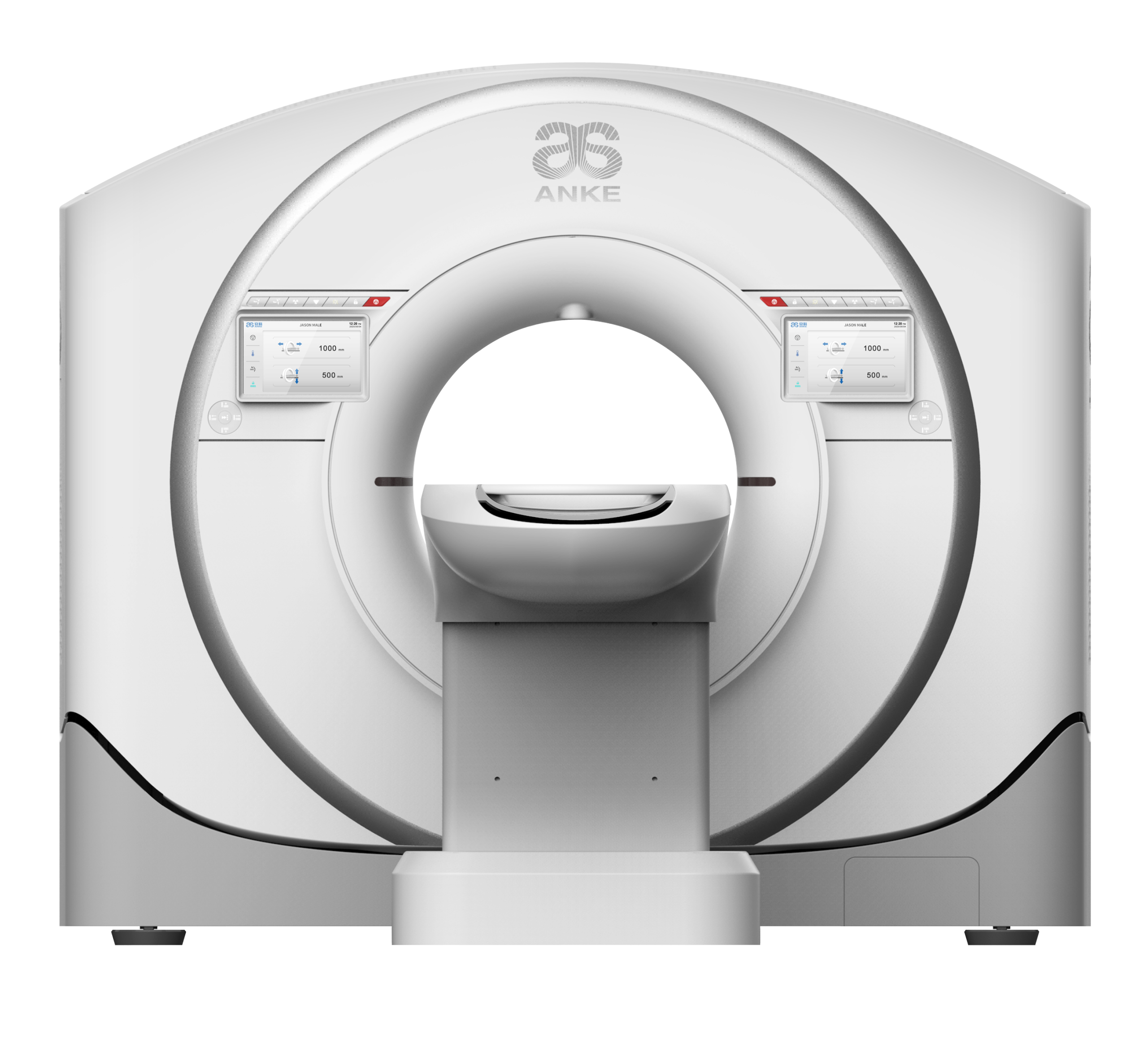
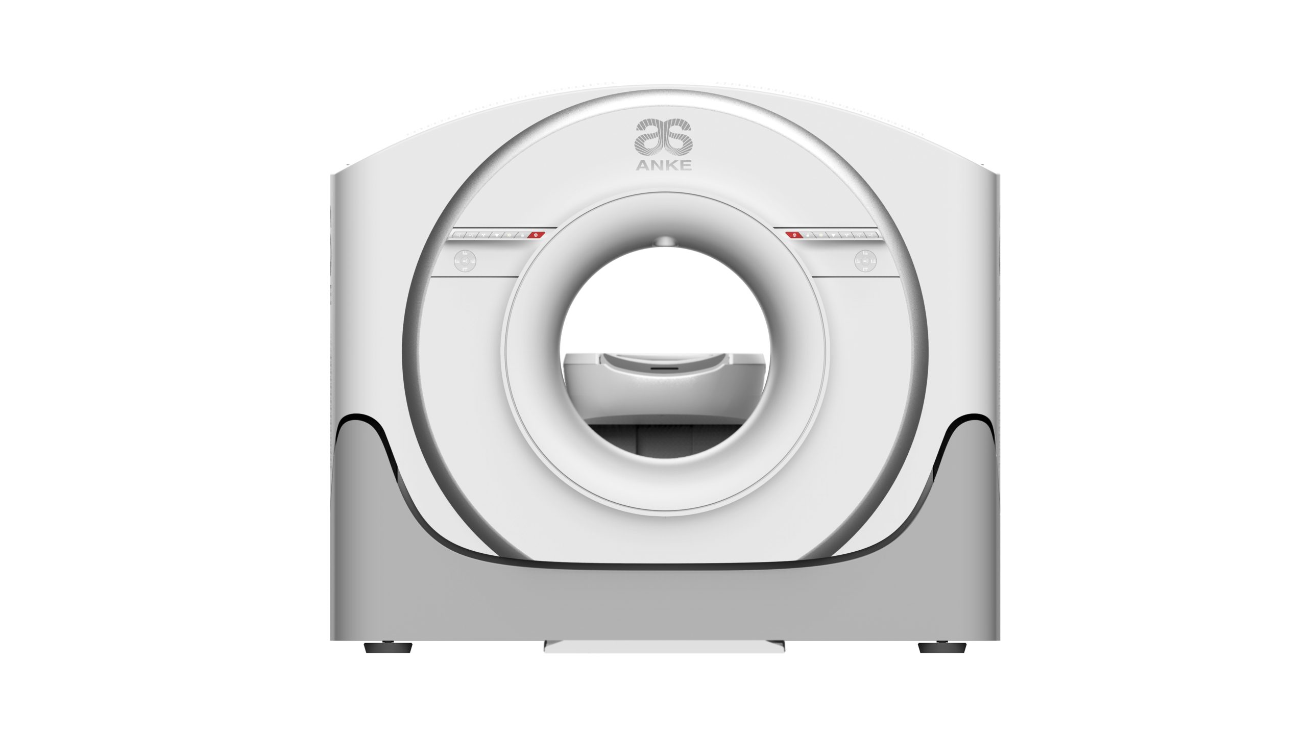
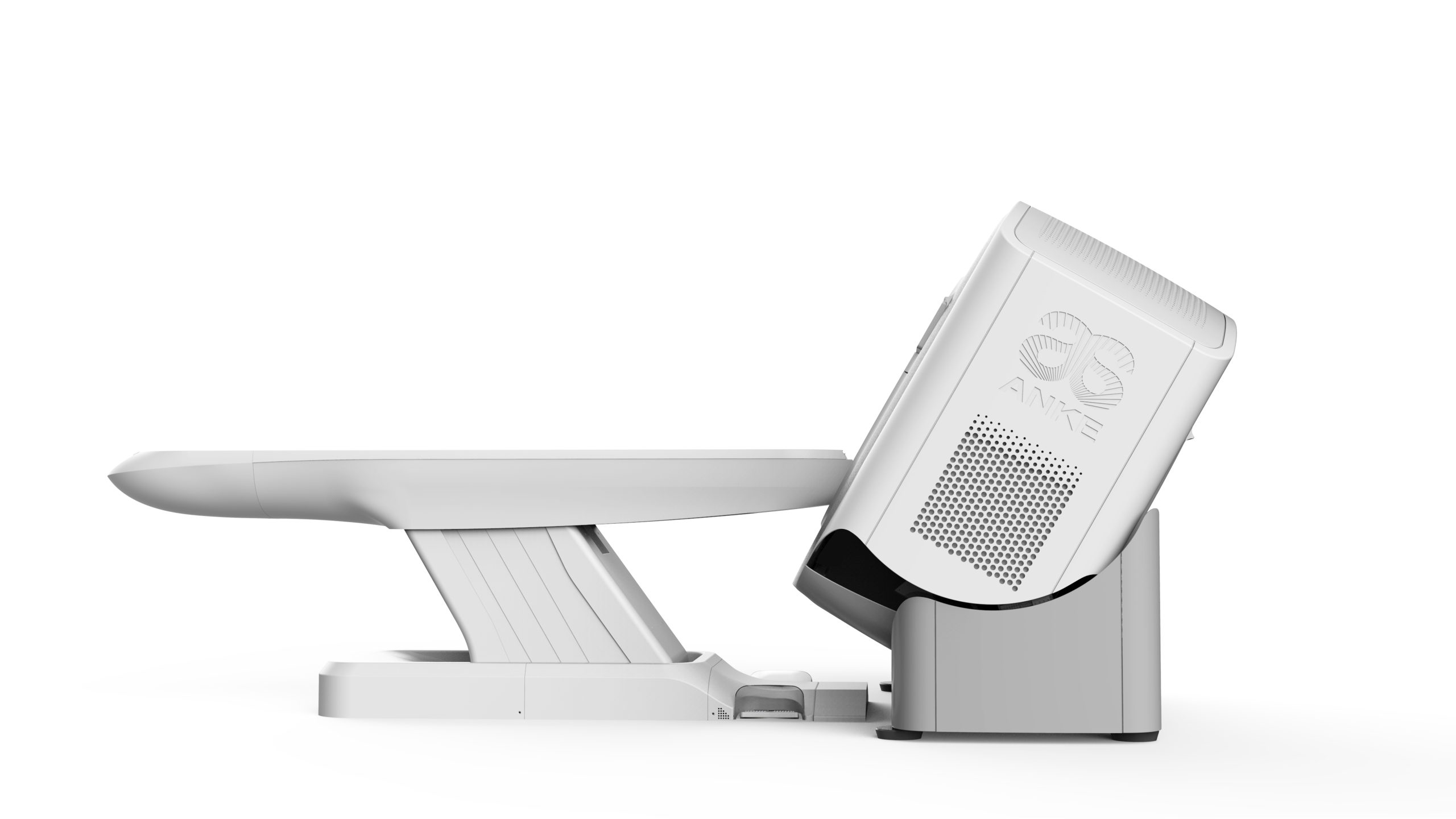
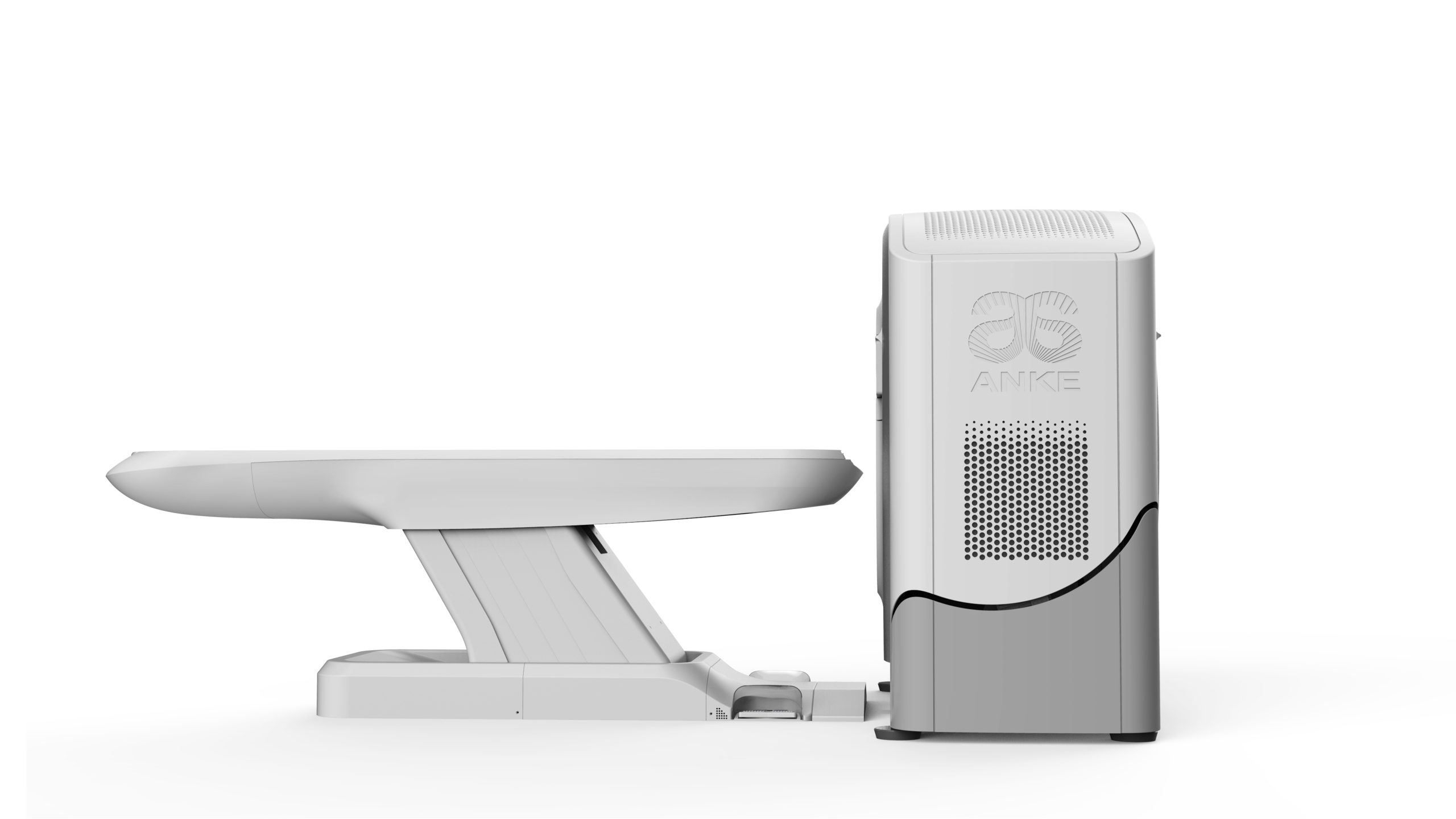
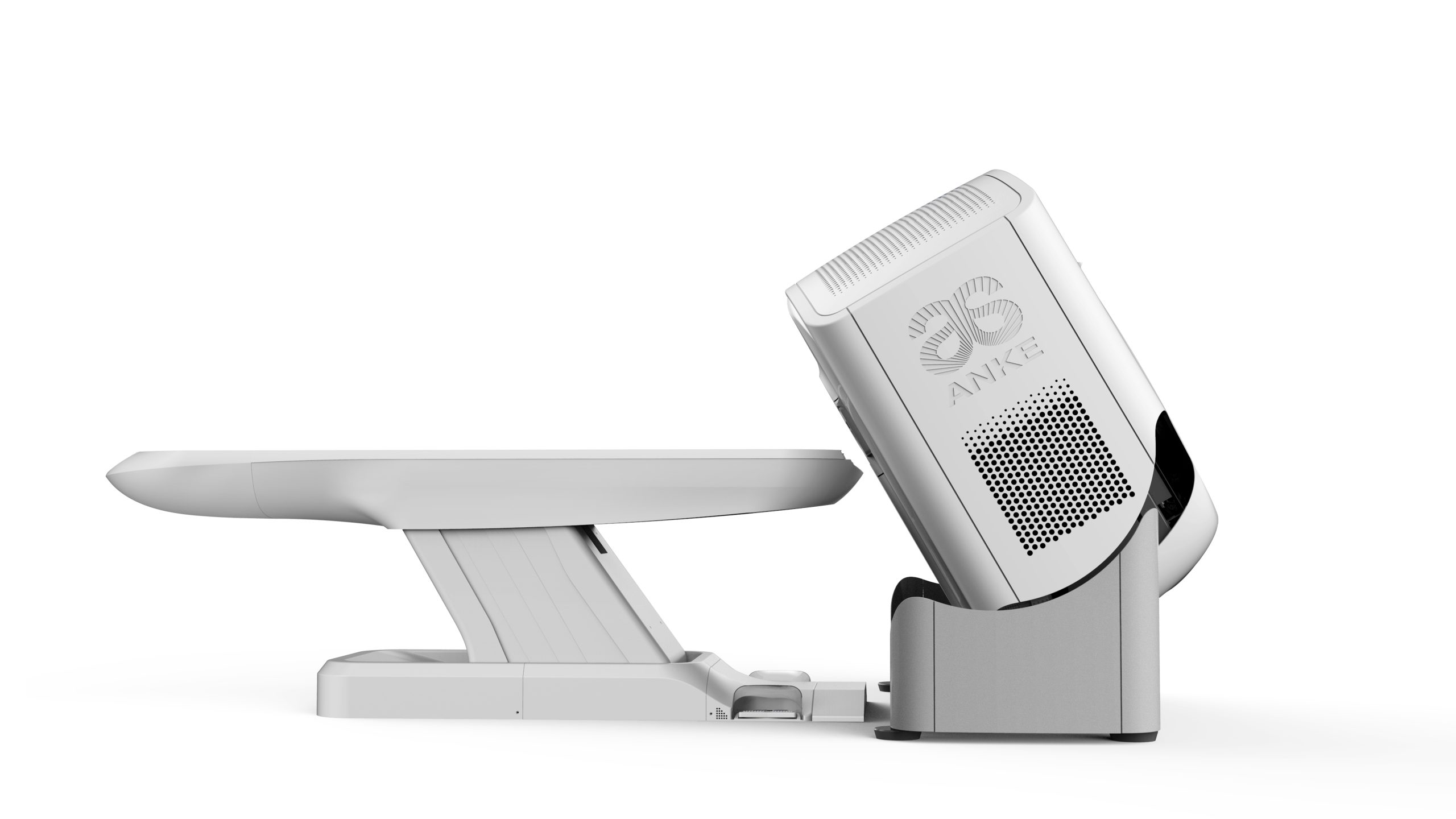
The ANATOM P428 X-ray computed tomography scanner is a modern computed tomography scanner with 64 physical slices per revolution and 128 reconstructed slices, which can be used for X-ray computed tomography of the head, trunk, heart and blood vessels and other parts of the body of patients of all ages. ANATOM P428 provides accurate and efficient diagnostics, combines advanced automated processing technologies with high performance, providing excellent image quality of anatomical structures with minimal radiation exposure to patients.
- Transferring images to the network
- Evaluation of dynamic volumetric brain scanning
- Reporting and printing software
- Filming
- Software package for general vascular analysis with selection and segmentation for standard measurements
- Print images on thermal film
- Cardiac visualization program with retrospective and prospective ECG synchronization
- Software for three-dimensional visualization of anatomical structures
- Software for 3D volume reconstruction and editing
- General vascular analysis software package for measuring vessel diameter
- Software for obtaining maximum intensity projections (MIP)
- Maximum and minimum intensity projections (MIP and minIP)
- Provide image archiving
- Vascular analysis software package with the ability to delete/edit bone structures
- Software for automatic bone removal, measurements – angles, density, dimensions
- Ethernet network support
- Software for real-time modulation of radiation load
- Remote communication with the scanning room
- Multiplanar reconstruction (MPR)
- Software instructions in Ukrainian
- Support for DICOM standards
- Software for visualization of cerebral perfusion
- Specialized workstation for image post-processing with medical monitor
- Availability of an ECG monitor
- Software for minimum intensity projections (minIP)
- Motorized table drive


